


 80 / 172

Abstracts of the 22
nd
National Congress of Digestive Diseases / Digestive and Liver Disease 48S2 (2016) e67–e231
e141
P.03.5
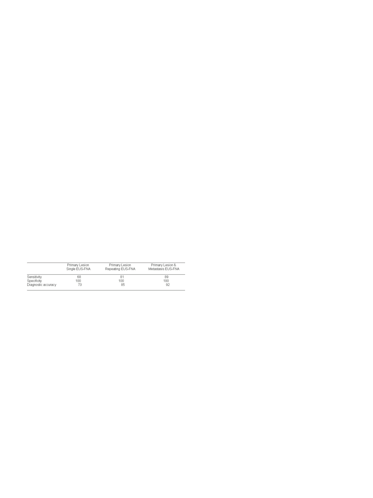
REPETITION OR SIMULTANEOUS SAMPLING OF PRIMARY AND
METASTATIC LESIONS IMPROVE DIAGNOSTIC ACCURACY OF
EUS-FNA IN THE ASSESSMENT OF SUSPECTED NEOPLASTIC
PANCREATIC MASS
Del Vecchio Blanco G.*, Paoluzi O.A., Mannisi E., Bevivino G.,
Formica V., Portarena I., Roselli M., Francesco P., Giovanni M.
University Tor Vergata, Roma, Italy
Background and aim:
Endoscopic ultrasound-fine needle aspiration
(EUS-FNA) is a relatively low invasive technique for diagnosing a
suspected neoplastic pancreatic mass. Several factors may influence
the adequacy of tissue collection leading to not conclusive findings.
Aim of the study was to evaluate if repetition or simultaneous
sampling of primary and metastatic lesions improve diagnostic
accuracy of EUS-FNA in patients with suspected pancreatic
malignancy.
Material and methods:
All patients with suspected malignancy
of the pancreas were submitted to EUS-FNA. In case of suspected
metastasis in the liver or lymph nodes, EUS-FNA of primary and
secondary lesions was performed in a same session. Final diagnosis
was defined according to surgical histopathology or clinical follow-
up.
Results:
A total of 126 patients (73 males, median age: 68 years,
range: 41-86) with a pancreatic mass underwent 142 EUS-FNAs:
102/126 patients (81%) with no evidence of metastasis underwent
EUS-FNA of pancreatic mass while in 24/126 patients (19%)
simultaneous EUS-FNA of primary lesion and metastases in the liver
(8 patients) or lymph nodes (16 patients) was performed. EUS-FNA
was repeated in 16/102 patients (15%) with no metastasis due to non
conclusive findings of first procedure. Both repetition of sampling or
simultaneous EUS-FNA of primary and metastatic lesions resulted to
improve sensitivity and diagnostic accuracy of the procedure (see
Table).
Conclusions:
Simultaneous sampling of primary lesion and
metastasis may increase EUS-FNA diagnostic accuracy and reduce
the need to repeat sampling due to non diagnostic findings in
patients with suspected neoplastic pancreatic mass.
P.03.6
COMPARING EUS-FNA AND ERCP-BRUSHING IN THE DIAGNOSTIC
WORKOUT OF SUSPECTED CHOLANGIOCARCINOMA: A
RETROSPECTIVE SINGLE-CENTER ANALYSIS
Dabizzi E.*, Testoni S.G., Occhipinti V., Petrone M.C., Mariani A.,
Arcidiacono P.G.
Pancreato-biliary Endoscopy and Endosonography Division, San
Raffaele Scientific Institute, Milan, Italy
Background and aim:
Cholangiocarcinoma is an aggressive tumor
and diagnosis still remains cumbersome.
Although novel intraductal techniques are emerging in the diagnostic
workout, cytology is still the gold standard. Diagnostic yield can also
depend on tumor location and characteristics as well as endoscopist
skill. EUS-FNA and ERCP brushing/biopsies are the most common
methods to obtain samples, although with conflicting data.
Data comparing these two techniques, for earlier diagnosis and
patient management are still lacking.
Aim of the study was to compare the diagnostic yield of EUS-FNA
vs ERCP brushing cytology in the diagnosis of cholangiocarcinoma
biliary strictures.
Material and methods:
A retrospective analysis was conducted
on the Endoscopy Database, from January 2013 and October 2015,
querying for patients undergone to EUS and/or ERCP for suspected
primary malignant biliary strictures. Patients demographics
and biliary ducts characteristics were recorded.Procedures were
performed under deep sedation, with anesthesiology assistance
by endoscopists expert in bilio-pancreatic procedures.Samples
were evaluated “on site” by expert cytotechnologist, after quick
hematoxilin eosin standardized staining for qualitative adequacy
and reviewed by an expert cyto-pathologist, for final diagnosis. Final
diagnosis was based on surgical pathology findings, where available,
and cythology. Lesions were divided into groups according to EUS
features and site. Data were analyzed with Student’s t-test and chi
squared test, assuming a significant p-value of 0.05.
Results:
62 patients (33M, mean age 70±9.8 years) underwent EUS
and/or ERCP during the study period. Out of these, in 51/62 pts (82%)
we reached a histo/cytological final diagnosis. In 45/51 pts (88%)
cholangiocarcinoma, in 2/51 pts (4%) pancreatic adenocarcinoma
and in 4/51pts (8%) other etiology were reported.
EUS-FNA diagnosed cholangiocarcinoma in 19/22 pts (sensibility
86%), whereas ERCP-brushing 23/36 pts (sensibility 64%)(p=0.06).
EUS-FNA was more sensitive and accurate than ERCP-brushing in
mass-forming lesions (100% vs 80%), intra-ductal vegetations (100%
vs 33%; p < 0.05), and duct wall thickness associated to solid nodules
(78% vs 59%), whereas ERCP-brushing was superior in case of duct
wall thickness (50% vs 0%).
Furthermore, in case of mass-forming cholangiocarcinoma, in which
both techniques were applied (5/22 pts; 23%), final diagnosis was
achieved only with EUS-FNA.
Conclusions:
In the diagnosis of cholangiocarcinoma EUS-FNA
is superior to ERCP-brushing in case of mass-forming lesions and
intra-ductal vegetations, although not statistically significant.
Depending on lesions characteristics, EUS-FNA should be considered
as first choice in the cholangiocarcinoma diagnostic armamentarium.
In case of ductal thickness ERCP-brushing is still superior.Further
prospective studies, also using novel endoductal techniques are
needed.
P.03.7
EUS-GUIDED FINE NEEDLE ASPIRATION OF SOLID PANCREATIC
TUMORS IN YOUNG PATIENTS: EXPERIENCE IN A TERTIARY
REFERRAL CENTER
Petrone M.C.*, Dabizzi E., Testoni S.G.G., Mariani A., Arcidiacono P.G.
San Raffaele Scientific Institute, Pancreato-Biliary Endoscopy and
Endosonography Division, Milan, Italy
Background and aim:
Pancreatic solid lesions in young patients are
relatively rare and, to our knowledge, the clinical value of pancreatic
masses fine needle aspiration (FNA) in patients < 40 years of age
remains limited.
Aim of this study was to evaluate the clinical value of EUS-FNA for
diagnostic evaluation of young patients with a pancreatic solid
lesion.
Material and methods:
A computerized search of our database
was performed for a period of 7 years. All pancreatic EUS-FNA cases
performed on patients less than 40 years of age were identified. Age,
gender, and the cytologic diagnosis were recorded for each patient
with pancreatic solid lesion. All available corresponding surgical
pathology reports or at least 6 months of follow-up were reviewed.
Results:
From October 2008 to October 2015, 3371 patients
underwent pancreatic EUS with FNA for solid or cystic pancreatic
tumor. Among these, 125 (3.7%) were aged between 11 and 40 age.