


 121 / 172

e182
Abstracts of the 22
nd
National Congress of Digestive Diseases / Digestive and Liver Disease 48S2 (2016) e67–e231
in the other two cases. Patients were found symptoms-free at
follow-up. One patient underwent a second stent placement.
Conclusions:
The present method results a safe alternative
endoscopic procedure in very selected patients when surgery is not
indicated.
P.11.2
ENDOSCOPIC ULTRASONOGRAPHY IN THE DIAGNOSIS AND
STAGING OF NEUROENDOCRINE TUMORS
Fugazza A.*, Cortegoso P., Gaiani F., Bizzarri B., De’ Angelis G.L.
AOU Parma, Parma, Italy
Background and aim:
Gastroenteropancreatic neuroendocrine
tumors (GEP-NETs) are nosological entities, whose incidence has
dramatically increased during the last decades.
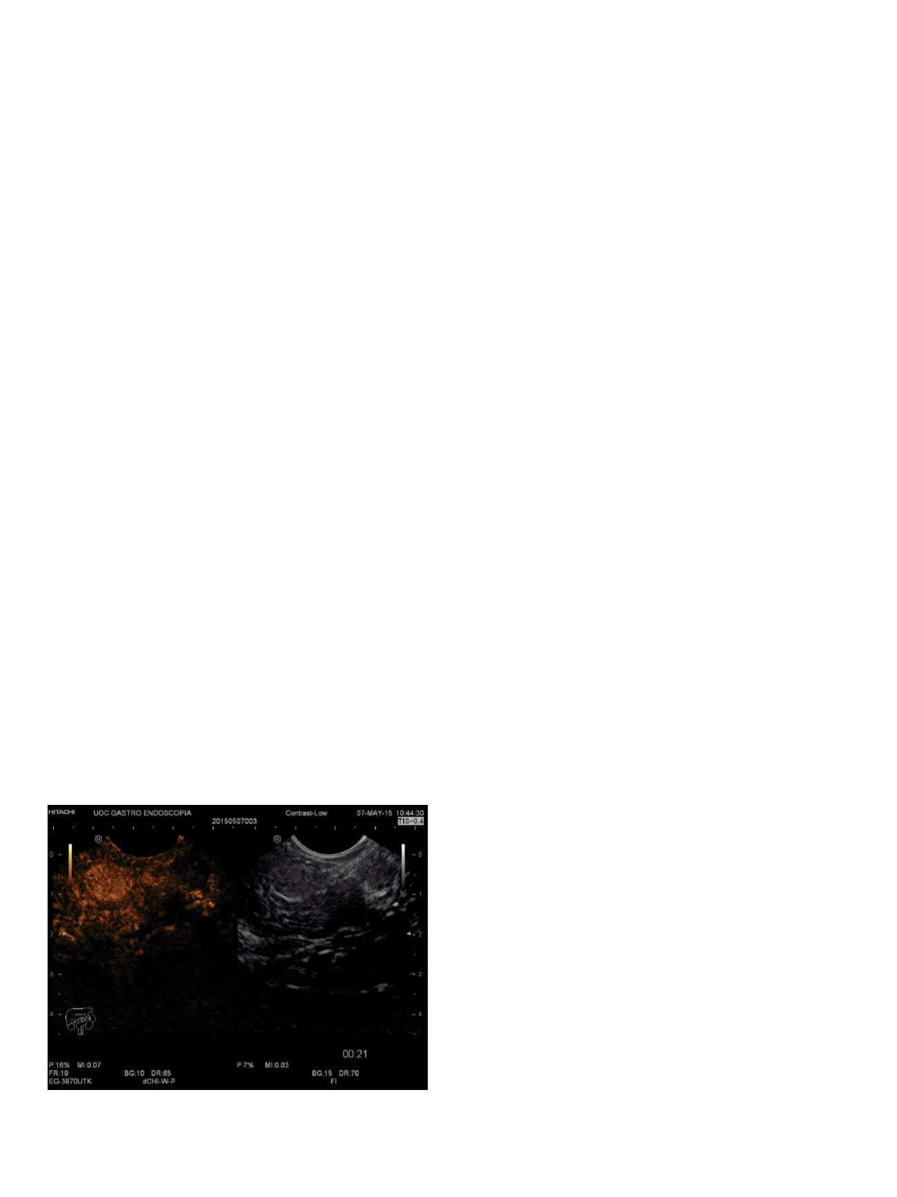
Endoscopic ultrasonography (EUS), associated to FNA and harmonic
contrast-enhancement (CH-EUS), has been reported to be extremely
useful for the diagnosis and the staging.
The objective of this study is to evaluate the accuracy of EUS in the
diagnosis and the staging of GEP-NETs.
Material and methods:
From January 2010 to September 2015, all
NET’s patients referred for EUS in our center were enrolled in this
study.
According to the localization of the tumor, the patients also
underwent laboratory tests and imaging techniques such as CT, MRI,
SRS Octreoscan or DOTATOC. EUS procedures were performed using
radial or linear echoendoscopes Pentax EG-3670URK–EG-3870UTK
(Pentax Hamburg, Germany) with a Hitachi – Aloka Avius processor
(Hitachi, Hamburg, Germany).
FNA procedures were performed with 25G FNA biopsy needles
(EchoTip, Wilson-Cook Medical Inc, Winston-Salem, NC) and
SonoVue (Bracco, Milano, Italy) was used for CH-EUS.
Results:
26 patients were enrolled in the study (17 m, 9 f) with
median age of 56.9 years (range 10 - 87).
NET’s were located in upper GI tract in 9 patients (6 stomach, 3
duodenum), in the rectum in 7 patients and in the pancreas in 10
patients.
In the patients with upper GI NET’s, found at bioptic sampling, EUS
confirmed endoscopical resection in 1 patient; surgical resection
in 4 patients because of an invasion of the deeper layers; medical
treatment in 4 patient with advanced disease.
In the patients with rectal NET’s, found at bioptic sampling in
colonoscopy, EUS permitted to choose the mucosectomy in 6
patients, and in 1 case surgical approach.
In pancreatic localization, CH-EUS showed a fast enhancement
with a homogeneous pattern lesion in 3 patients with recurrent
hypoglycemias. FNA confirmed the diagnosis of insulinoma in all
cases.
CT suspected a pancreatic NET in the other 7 patients, EUS+FNA
confirmed the presence of neuroendocrine tumor. FNA was
performed with a mean of 2.0 passages per patient.
Three patients underwent surgery, while the others underwent
medical therapy for the advanced disease.
Neither major or minor complications showed up during or after the
procedures.
Conclusions:
This study highlights the diagnostic accuracy, safety of
EUS in the evaluation and management of GEP-NETs. In particular,
EUS was necessary to define whether the lesion could be managed
endoscopically or surgically.
P.11.3
THE ROLE OF COMBINED USE OF EUS-FNA AND BILIARY
BRUSHING IN CYTOLOGICAL DIAGNOSIS OF PANCREATOBILIARY
MALIGNANCES
Bulajic M.*
1
, Vieceli F.
1
, Berretti D.
1
, Zoratti L.M.
1
,
Vadala’ Di Prampero S.F.
1
, Marino M.
1
, Toso F.
1
, Panic N.
2
, Terrosu G.
1
,
Zilli M.
1
1
Academic Hospital “S. M. della Misericordia”, Udine, Italy,
2
University
Clinic “Dr Dragisa Misovic-Dedinje”, Belgrade, Serbia
Background and aim:
Fifteen percent of patients with suspected
pancreatobiliary malignancy that undergo surgery without
a cytological assessment have a benign lesion. Cytological or
histological diagnosis of pancreatobiliary malignancies before
surgery is desirable in order to avoid unnecessary interventions.
We conducted a study in order to assess whether the combined use
of biliary brushing and endoscopic ultrasound-guided fine needle
aspiration (EUS--FNA) has greater accuracy than the individual
procedures in diagnosing pancreatobiliary malignancies.
Material andmethods:
Studywas conducted at theGastroenterology
Unit of Academic Hospital “S. M. della Misericordia”, Udine, Italy.
Twenty five patients with probable pancreatobiliary malignancy
were subjected both to biliary brushing and EUS-FNA and collected
material was sent for cytological analysis. The results of cytology
were compared to the results of histology from surgical specimen.
Results:
Histology of surgical specimen confirmed the diagnosis
of pancreatobiliary malignancy in 24 of 25 patients, benign lesion
caused by chronic pancreatitis was identified in one patient.
Cytology from biliary brushing provided a correct diagnosis in
9 patients, with diagnostic accuracy of 36%. For the remaining 16
patients (54%), cytological diagnoses were as follows: indeterminate
because of poor quantity or quality of the specimen in 15 patients,
negative in one case (1 false negative). EUS-FNA provided a correct
diagnosis in 18 patients with diagnostic accuracy of 72%, including
the patient with a benign lesion. In 7 patients (28%) EUS-FNA didn’t
provide any result because of the poor quality of the specimen. The
combined diagnostic accuracy of both methods was 80% as they
together provided a correct diagnosis in 20 patients. The additional
diagnostic gain derived from the joint use of biliary brushing and
EUS-FNA was +44% compared to biliary brushing alone and +8%.
cases compared to EUS-FNA alone.
Conclusions:
The combined use of the EUS-FNA and biliary
brushing results in increased accuracy of cytological diagnosing
in pancreatobiliary malignancies. The biliary brushing as an
addition to EUS-FNA should be considered in patients undergoing
the endoscopic retrograde cholangiopancreatography (ERCP) in
therapeutic purposes.