


 139 / 172

e200
Abstracts of the 22
nd
National Congress of Digestive Diseases / Digestive and Liver Disease 48S2 (2016) e67–e231
was to evaluate clinical efficacy and safety of IFX and ADA in a single
IBD center in a real-life study.
Material and methods:
Clinical records of CD patients, that
underwent anti-TNF therapy (who started from January 2011 to
October 2014 and performed at least 3 infusions) at S. Andrea
Hospital in Rome, Italy, were retrospectively collected. Baseline
characteristics of patients before the start of biologic therapy
were recorded. For the biologic therapy, the duration, side effects,
and interruption rate, were considered. Finally, clinical remission
(defined as the complete absence of clinical symptoms) and clinical
response (amelioration of clinical symptoms from baseline) rate, at
1 year of follow-up, in intention-to-treat (ITT) and per protocol (PP)
analysis, was recorded.
Results:
A total of 30 patients (16 treated with IFX and 14 with ADA)
were included. Baseline characteristics between the two groups did
not differ except for the rate of biolologic naïve patients, that was
significantly higher in IFX than in ADA group (94% vs. 57%, p<0.05).
Four patients stopped the therapy before 1 year, two in IFX (1 primary
non response, 1 prostate cancer diagnosis) and two in ADA group
(1 death unrelated to therapy, 1 extensive psoriasis onset). Adverse
events that require postponement of therapy schedule occurred in 5
(31%) and 2 (14%) of patients in IFX and ADA group, respectively. At 1
year, clinical remission was observed in 50% (44% and 57% in IFX and
ADA group) and clinical response in 70% of patients (63% and 79%
in IFX and ADA group, respectively) at the ITT analysis, and in 58%
(50% and 67% in IFX and ADA) and 81% (71% and 92% in IFX and ADA
group, respectively) at the PP analysis.
Conclusions:
The present retrospective single center real-life study
confirms that infliximab and adalimumab are safe and effective
therapies in CD patients. Prospective multi-center head-to-head
studies would better clarify specific profile of the two anti-TNF
a
drugs.
P.14.16
A NEW THERAPEUTIC LASER SYSTEM FOR ENDOSCOPIC
ABLATION OF ESOPHAGEAL LESIONS – FIRST RESULTS IN AN
ESTABLISHED ANIMAL MODEL
Tontini G.E.*
1
, Soriani P.
1
, Neumann H.
2
, Spina L.
1
, Annunziata M.L.
1
,
Vavassori S.
1
, Fagnani F.
3
, Carmignani L.
4
, Pastorelli L.
1
, Vecchi M.
1
1
Gastroenterology & Digestive Endoscopy Unit, IRCCS Policlinico San
Donato, San Donato Milanese, Milano, Italy,
2
Department of Medicine
I, University of Erlangen-Nuremberg, Erlangen, Germany,
3
Surgical
Division, Quanta System S.p.A, Varese, Italy,
4
Academic Urology
Department, IRCCS Policlinico San Donato, San Donato Milanese,
Milano, Italy
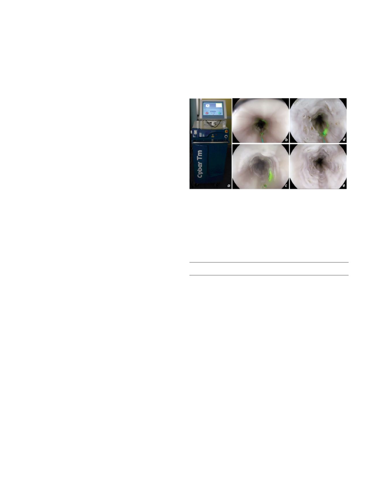
Backgroundandaim:
TheThuliumlaser systemis anovel therapeutic
technique for open surgery and endourological treatments [1; fig.
a]. To date, the experience on the use of this therapeutic device
in gastrointestinal (GI) endoscopy is very limited [2]. Recent
experience on animal models showed that the wavelength of 2μm
allows for ablation and vaporesection of the superficial GI layer
providing high control on penetration depth (0.2-0.4 mm) and tissue
damage [3]. We conducted a pilot study in an established animal
model (EASIE) to test for the first time both feasibility and safety of
the Thulium Laser system (Cyber TM®, Quanta System, Varese, Italy)
for endoscopic ablation of pre-neoplastic esophageal lesions, such
as Barrett esophagus.
Material and methods:
According to previous experience [3] and
the need of lateral ablative effect, we used a dedicated 600 um
side fiber with a line beam that emerges at 45 degrees with soft
power settings (5-10 watts) and continued laser modality. For
safety, the study endpoints was the impact of the laser ablation in
terms of depth penetration and lateral tissue damage after having
vaporesected circumpherentially a 3 cm-length luminal esophageal
surface. All procedures were performed using a high-definition
video-gastroscope and digitally video-recorded.
Results:
Neither transmural perforation, nor any submucosal layer
damagewas observed. Two endoscopists completeda circumferential
ablation of a 3cm-length luminal esophageal surface within 1
minute each. Overall, each laser ablation on target produced mucosal
vaporesection with only a diminutive lateral spreading of epithelial
injury (1-3 mm), depending on the distance between the fiber’s tip
and the esophageal target (fig. b-e).
Conclusions:
The Thulium laser system appears to be safe, effective,
and very easy to use for the ablation of superficial esophageal lesions
in an ex vivo animal model. In vivo studies should now confirm these
initial results in a prospective setting.
References:
1. Rieken M & Bachmann. Nat Rev Urol 2014.
2. Cho JH, et al. Endoscopy 2013.
3. Tontini GE, et al. UEG Week 2015.
P.15 Endoscopy 1
P.15.1
ENDOSCOPIC RESECTION OF DUODENAL NEUROENDOCRINE
TUMORS: A CASE SERIES OF A SINGLE INSTITUTION
Fiori G.*
1
, Ravizza D.
1
, Trovato C.
1
, De Roberto G.
1
, Bravi I.
1
, Genco C.
1
,
Bottiglieri L.
2
, Crosta C.
1
1
Division of Endoscopy, European Institute of Oncology., Milan, Italy,
2
Division of Pathology, European Institute of Oncology., Milano, Italy
Background and aim:
Duodenal neuroendocrine tumors (dNETs)
represent 1-3% of all primary duodenal tumors. dNETs not located
in the periampullary region are suitable for endoscopic treatment
if limited to the submucosal layer, without metastases and with
size <10 mm. Nevertheless, few data are available about the efficacy
of this approach. We reviewed our data about dNETs treated with
endoscopic resection (ER).
Material andmethods:
From 2012 to 2014, 11 dNETs were diagnosed
during upper GI endoscopy (UGIE). Five of them were considered
suitable for ER. The endoscopic procedure was performed with a
high definition single-channel endoscope (EG29i series, Pentax)
and the resection technique was chosen according to endoscopist’s
preference, morphological characteristics, site and endosonographic
features of each lesion.
Results:
En-bloc resection was obtained in all cases. Endoscopic
mucosal resection (EMR) was used for 3 bulb tumors and endoscopic
submucosal dissection (ESD) for one bulb tumor. One tumor of distal
duodenum was treated with Hybrid-ESD (HESD). In 4 cases the
resection site was closed with metal clips. The mean size of resected
specimens was 14 mm (range 7-22 mm); histologically the tumor