


 60 / 172

Abstracts of the 22
nd
National Congress of Digestive Diseases / Digestive and Liver Disease 48S2 (2016) e67–e231
e121
Material and methods:
We report our experience with urgent
Ercp (defined as a procedure performed within 12 hours after
clinical presentation) at our institution in the period January 2013
- September 2015.
The indication to urgent Ercp was severe pain associated with
biliary obstruction, with imaging (computerized tomography scan,
magnetic resonance, endosonography) evidence of stone impaction
in the papilla.
The series consisted of 7 patients, 4 males; mean age 46 years, range
19-92.
Results:
In 4 cases an urgent Ercp has been performed, with fast and
satisfactory resolution of the clinical picture and no complication
observed; in three this has not been possible and the procedure
has been executed 2-3 days later: in two cases pain has been very
difficult to control and in one patient an acute pancreatitis has
developed.
Conclusions:
Our experience suggests to make all efforts to perform
an early Ercp in patients with stone impaction in the papilla, due
to possible development of intractable pain and unpreventable
complications.
V.01.7
ENDOSCOPIC ULTRASOUND-GUIDED SINGLE-INCISION WITH
NEEDLE KNIFE AND DEEP TISSUE BIOPSY FOR THE DIAGNOSIS OF
A GASTRIC SUBEPITHELIAL TUMOR
Antonini F.*
1
, Belfiori V.
1
, Santinelli A.
2
, De Minicis S.
1
, Lo Cascio M.
1
,
Marraccini B.
1
, Piergallini S.
1
, Rossetti P.
1
, Andrenacci E.
1
, Macarri G.
1
1
Ospedale A.Murri, Fermo, Italy,
2
Ospedali Riuniti, Ancona, Italy
Background and aim:
Gastrointestinal subepithelial tumors (SETs)
includes a variety of neoplastic and non-neoplastic lesions that can
be difficult to diagnose. Endoscopic ultrasound (EUS) is currently
recommended as a first choice for examining SETs, even if its
diagnostic yield seems to be suboptimal. Therefore, several other
techniques for sampling SETs have been utilized.
Material and methods:
An 80-year-old man was referred to our
unit for the evaluation of a gastric SET. An EUS revealed a 25 mm
homogenous hypoechoic well-circumscribed tumor, originating
frommuscular layer. An EUS-fine needle biopsy of the lesion resulted
inconclusive. Therefore a EUS-guided single-incision with needle
knife (EUS-SINK) biopsywas performedusing a linear echoendoscope
guiding a 10-mm linear incision over the lesion through a needle-
knife sphincterotome connected to an electrosurgical unit. Then a
conventional biopsy forceps were introduced to obtain deep tissue
samples. Subsequently, the incision was closed with an endoclip.
Procedure was uneventful.
Results:
Histology showed a group of spindled-shaped cells resulted
positive for CD117 and DOG-1 while negative for desmin, smooth
muscle actin and S-100 expression on immunohistochemistry,
in keeping with a gastrointestinal stromal tumor. The patient
underwent surgical resection.
Conclusions:
In this article we report on a more accurate diagnostic
possibility offered by EUS-SINK with deep tissue biopsy for
pathologic diagnosis of a gastric SET.
V.01.8
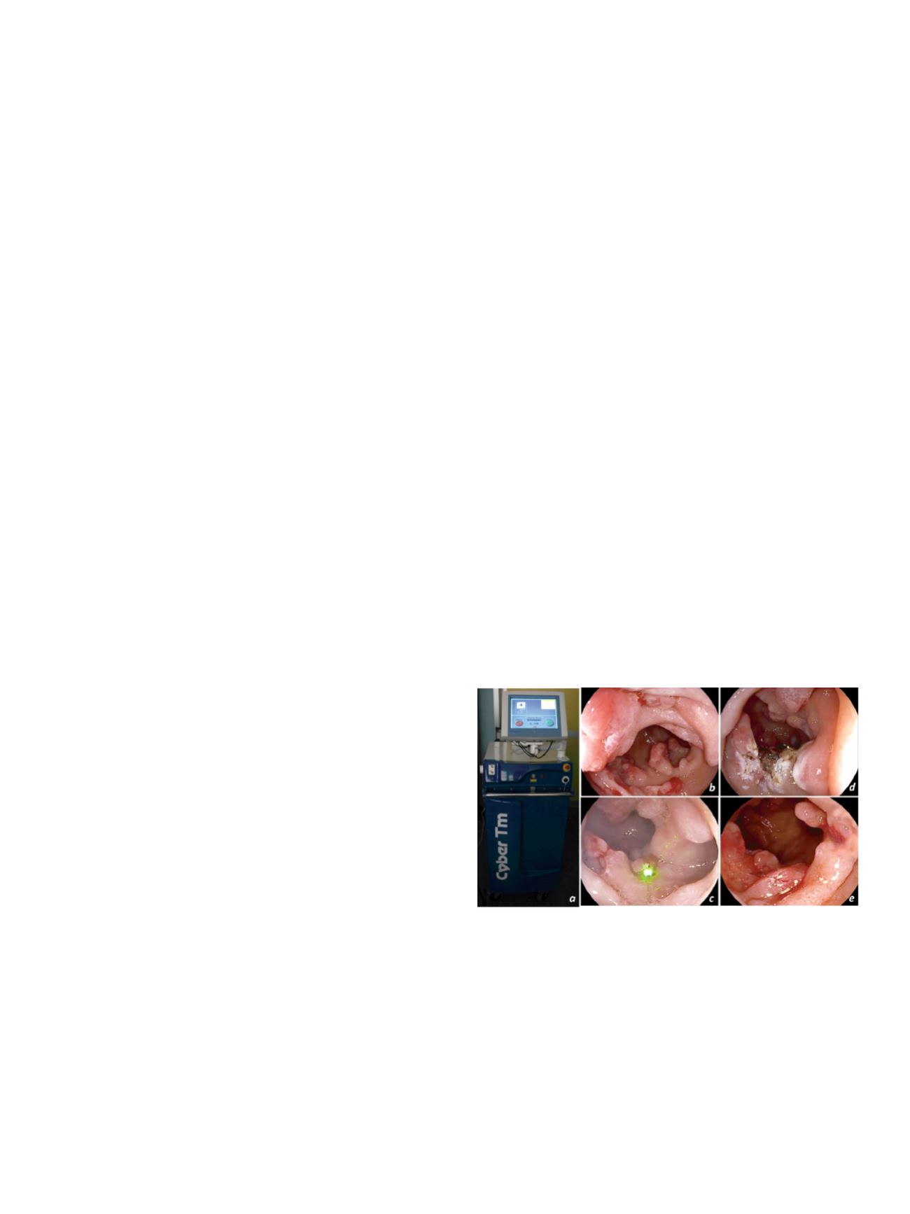
HAEMOSTATIC TREATMENT WITH A NEW THERAPEUTIC LASER
SYSTEM – FIRST IN VIVO EXPERIENCE (WITH VIDEO)
Tontini G.E.*
1
, Soriani P.
1
, Neumann H.
2
, Carmignani L.
3
, Fagnani F.
4
,
Spina L.
1
, Annunziata M.L.
1
, Vavassori S.
1
, Pastorelli L.
1
, Vecchi M.
1
1
Gastroenterology & Digestive Endoscopy Unit, IRCCS Policlinico San
Donato, San Donato Milanese, Milano, Italy,
2
Department of Medicine
I, University of Erlangen-Nuremberg, Erlangen, Germany,
3
Academic
Urology Department, IRCCS Policlinico San Donato, San Donato
Milanese, Milano, Italy,
4
Surgical Division, Quanta System S.p.A,
Varese, Italy
Background and aim:
The Thulium laser system (fig. a) is an
established therapeutic technology for surgical resection [1]. By
adjusting the power, its wavelength of 2μmprovides a precise control
on penetration depth (0.2-0.4mm) for ablation and vaporesection
purposes in luminal endoscopy [2]. Here, we report on the first in
vivo haemostatic treatment in humans, with this newly introduced
tool during ongoing gastrointestinal bleeding, which had not been
controlled by means of conventional haemostatic methods.
Material and methods:
The new therapeutic laser system was
used in a 67-year old man with recurrent oozing duodenal bleeding
from a large post-inflammatory elevated lesion placed along the
proximal duodenum. One year before, the patient had undergone a
rescue treatment with selective arterial embolization for persistent
active bleeding despite several endoscopic attempts in a huge and
deep peptic ulcer located at the same part of the duodenum. The
patient developed a large post-inflammatory duodenal lesion with
recurrent oozing bleedings, which were unsatisfactorily controlled
by standard thermal, cytochemical, and mechanical devices [3]. The
endoscopic examination was performed using a high-definition
videogastroscope and digitally video-recorded.
Results:
Under conscious sedation, the endoscope was advanced
into the duodenum, thereby showing two areas of oozing bleeding
within the post-inflammatory lesion (fig. b). Then, a 550 um optical
fiber was introduced into the working channel, placing the tip at a
distance of approximately 1 cm from the endoscope and from each
targets. Using an integrated green laser as a pilot light for tissue
targeting (fig. c), the Thulium laser system was used as a paintbrush
to carefully vaporise the mucosal surface under a 5 watts continued
modality. When active bleeding from an exposed vessel occurred,
the focal administration of 10 watts power resulted in an immediate
and persistent haemostatic ablation (fig. d). Patient was discharged
home 4 hours after the procedure and no adverse event was
recorded. Four weeks later, the endoscopic control revealed an
initial mucosal healing upon the targeted area (fig. e).
Conclusions:
The Thulium laser system appears to be safe and
effective for in vivo haemostatic therapy of active bleeding lesions
in the upper GI-tract, which are not amendable with conventionally
haemostatic therapies. Multicenter studies should now confirm
these initial results in a prospective setting.
References
:
1. Rieken M & Bachmann A. Nat Rev Urol 2014.
2. Tontini GE, et al. UEG Week 2015.
3. ASGE Technology Committee. Endoscopic hemostatic devices. Gastrointest
Endosc 2009.