


 67 / 172

e128
Abstracts of the 22
nd
National Congress of Digestive Diseases / Digestive and Liver Disease 48S2 (2016) e67–e231
We report a case of endoscopic drainage of a pancreatic pseudocyst
through a gastro-gastro-cyst anastomosis in a patient who
underwent a laparoscopic Roux-en-Y gastric bypass for obesity.
Material and methods:
A 33-year-old female with Roux-en-Y
gastric bypass was admitted to our hospital because of a CT evidence
of symptomatic 7 cm pancreatic cystic lesion. Laboratory indicated
iron deficiency anaemia. An endoscopic ultrasound (EUS) evaluation
was performed. From gastric stump a cystic lesion of 7 cm in size
was observed, but the excluded gastric pouch was interposed. A fine
needle aspiration with a 19G needle (ECHO-19, Cook Medical) was
performed. The cytological analysis showed granulocytes, histiocytes
and was negative for malignant cells. Amylase and CEA levels were
respectively 6785 U/ml and <5 ng/ml. Then we proposed an
endoscopic approach. Initially an EUS-guided puncture from the
gastric stump with a 19 G needle was performed and an access to
excluded gastric lumen was obtained; after injection of contrast
medium, a 0.035-guidewire was then placed into the excluded
gastric pouch, and a gastrogastric fistula was created by pushing a
10Fr cystoenterostome (XS 1341, Endoflex) on the guidewire. Finally,
a 10Fr-20mm, SEMS (Nagi stent; Taewoong Medical) was left in
place After 2weeks, failing to go trough the gastrogastric anastomosis
with a therapeutic echoendoscope (Pentax), SEMS was substituted
by a 20Fr-60mm enteral fully covered SEMS (Teawoong Medical).
One month later was possible to reach the excluded gastric pouch
with a therapeutic echoendoscope (Pentax) passing trough the
enteral stent. Then an EUS guided puncture from the gastric pouch
with a 19-gauge needle was achieved and a 0.035-guidewire was
placed into the cyst; a gastrocystic fistula was created by pushing a
10Fr cystoenterostome on the guidewire. Finally a 16 Fr -20mm,
SEMS (Nagi stent; Taewoong Medical) was left in place. Passage of
air in the peritoneal cavity occurred, It was evacuated by placement
a XX needle under CT guidance. The patient was discharged 72 h
later healtly Two months later CT showed complete drainage of the
cyst.
Results:
Two months later cross sectional imaging showed complete
drainage of the cyst.
Conclusions:
In selected cases and in experienced hands, EUS
guided drainage of pancreatic pseudocysts is a viable therapeutic
alternative also in patients with previous digestive surgery.
V.02.12
PERORAL CHOLANGIOSCOPY VIA SPYGLASS SYSTEM FOR
INDETERMINATE BILIARY STRICTURES: AN EFFECTIVE AND SAFE
TOOL TO DISTINGUISH MALIGNANT FROM BENIGN LESIONS
WHEN CONVENTIONAL METHODS HAVE FAILED
Sica M.*, Manta R., Tringali A., Mutignani M.
Surgical Digestive Diagnostic and Interventional Endoscopy, “Niguarda
Ca’ Granda Hospital”, Milano, Italy
Background and aim:
Diagnosing malignant etiologies of biliary
strictures is a difficult challenge. ERCP cytologic or tissue diagnosis
with brushing, biopsies, or both is limited by their poor sensitivity.
Peroral Cholangioscopy (POC) via the SpyGlass cholangioscopy
system (Spyglass®) is a safe and effective adjunctive tool with ERCP
for evaluation of bile duct strictures when conventional methods
have failed. We report a video-case of an indeterminate hilar biliary
stricture in whom SpyGlass was used for diagnostic purpose.
Material and methods:
A 71-year-old man with a recent history
of jaundice and weight loss (about 6 kg) and CT scan evidence of a
“mass forming” hilar biliary strictures, already underwent in another
hospital to PTBD, exploratory laparotomy and cholecystectomy with
inconclusive biopsy on the hilar mass, was admitted to our hospital
because of recurrent cholangitis.
A new CT scan showed increase in the size of the mass.
An EUS with FNA was performed but citological sample was not
representative.
CPRE showed the presence of proximal third bile duct stricture.
Cyto-histological sampling was performed by brushing and biopsies,
and were placed two plastic stents.
Since, cyto-histological examination was negative for malignant
cells, it was decided to refer the patient to Peroral Cholangioscopy
(POC) via the SpyGlass cholangioscopy system (Spyglass®) (VIDEO).
Results:
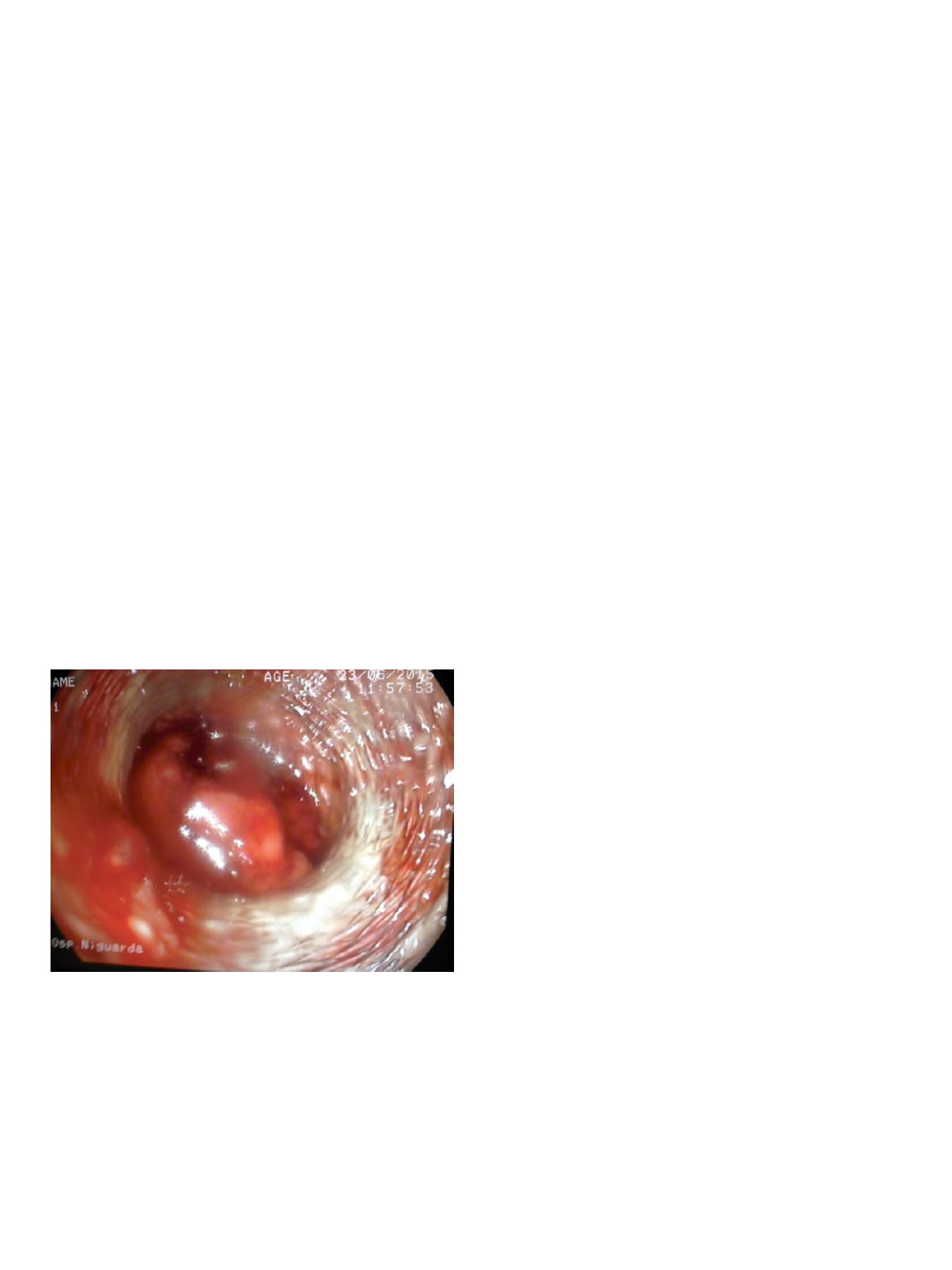
Direct visualization of the stenosis showed irregular
nodulations with erosions but no clear signs of malignancy
(Intraductal nodular/villosus masses; oozing and irregular vascular
patterns with an irregular surface). Histological examination
showing chronic inflammation without mlignant cells, allowed us
to exclude the presence of malignancy.
Conclusions:
Peroral Cholangioscopy (POC) via the SpyGlass
cholangioscopy system (Spyglass®) provides direct visualization
of strictures and allows for targeted biopsies, which may help
to diagnose or rule out malignancy in indeterminate strictures.
Future trials should develop algorithmic approaches incorporating
cholangioscopy targeted biopsies and validate them in diagnosing
patients with indeterminate biliary strictures.
V.02.13
METASTATIC MELANOMA OF THE GALLBLADDER DIAGNOSED BY
ENDOSCOPIC ULTRASOUND-GUIDED FINE NEEDLE BIOPSY
Antonini F.*
1
, Acito L.
1
, Sisti S.
2
, Angelelli L.
3
, Belfiori V.
1
,
De Minicis S.
1
, Lo Cascio M.
1
, Marraccini B.
1
, Piergallini S.
1
,
Rossetti P.
1
, Andrenacci E.
1
, Macarri G.
1
1
Ospedale A.Murri, Fermo, Italy,
2
Ospedali Riuniti Torrette, Ancona,
Italy,
3
Ospedale Mazzoni, Ascoli Piceno, Italy
Background and aim:
A 73 year-old woman with a history of
malignant cutaneous melanoma (BRAF wild type) of the groin
excised four years before, was referred for further characterization
of an asymptomatic gallbladder mass discovered during follow-
up on abdominal US then also detected on CT scan. Blood tests
showed mild elevation of gamma glutamyltransferase, erythrocyte
sedimentation rate and carcinoembryonic antigen. Endoscopic
ultrasound (EUS) confirmed a 30 mm irregular mass rising from the
gallbladder wall and extending into the lumen.